
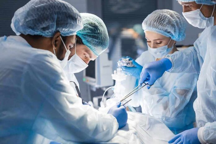
Surgeons are now literally making lung tumors glow in the operating room, and it is changing how cancer is removed.
Story Snapshot
- A fluorescent drug called pafolacianine makes certain lung nodules light up during surgery
- Doctors are finding hidden cancers that scans and touch alone would have missed
- The extra “glowing” lesions often sit outside the planned cut, forcing smarter course changes
- Early data show no serious drug-related side effects, but long-term impact still needs tracking
Turning lung cancer surgery into a guided light show
Pafolacianine is a targeted imaging drug that binds to lung cancer cells and glows when hit with near-infrared light during surgery. Surgeons inject it before the operation, then use a special camera in the operating room to see fluorescent nodules that look normal under regular white light. In a major phase 3 study known as ELUCIDATE, this glow helped surgeons find cancer lesions that their eyes and hands alone could not detect.
In that trial, intraoperative molecular imaging with pafolacianine located the main lung nodule that the surgeon could not find with white light and palpation in 19 out of 100 patients. Put simply, in almost one out of five cases, the “index” tumor hid from standard technique but lit up when the drug was used. The same study found additional malignant lesions in eight patients that were completely missed on preoperative imaging and standard inspection.
Changing the game while the patient is on the table
Lighting up these nodules does more than satisfy a surgeon’s curiosity; it changes the plan mid-surgery. In the ELUCIDATE trial, the scope of the operation shifted in 29% of patients once surgeons saw what was glowing. Some cuts got bigger to include cancer that would have been left behind. Some got smaller to preserve healthy lung tissue where no fluorescent signal appeared. This kind of real-time adjustment lines up with conservative values: get the job done right the first time and avoid waste.
Most of the surprise malignant lesions discovered with imaging were not where surgeons expected them. About 73% of the extra cancers found by intraoperative molecular imaging sat outside the original planned resection field. That means standard scans and logic would have missed them. For a patient, that can be the difference between a truly clean operation and a quiet leftover tumor that grows later. For surgeons, the drug acts like a flashlight showing the edges of the problem instead of guessing in the dark.
From trial results to real-world lung cancer practice
The United States Food and Drug Administration (FDA) reviewed this data and approved pafolacianine, sold as Cytalux, to help identify lung lesions during surgery in adults with known or suspected lung cancer. In the FDA’s own summary, 24% of patients had their main lesion and or extra cancerous lesions detected that were not seen by standard visual and tactile inspection. That is not hype from a company press release; it is a federal regulator saying the drug reveals disease that would otherwise stay hidden.
Real-world reports after approval support the trial findings. Surgeons using pafolacianine-based intraoperative molecular imaging describe “clinically significant events” where the glowing nodules force meaningful changes in how they operate. The most common event is localization of lesions that cannot be seen with white light cameras or felt through minimally invasive instruments. These same reports show higher impact during smaller, sublobar resections, where preserving lung tissue is a priority but the risk of missing something at the edge is higher.
Safety, limits, and the pattern of new imaging tools
Any new cancer tool raises a fair question: is this safe, and is the glow trustworthy. Safety data from the ELUCIDATE trial and follow-up experience in more than 700 patients show no serious adverse effects linked to pafolacianine. That matters for older adults already juggling other health issues. If a drug adds no major harm but helps find extra cancer, that is a strong practical trade-off many patients and doctors will accept without hesitation.
The glow is not magic, though. Studies show that the drug’s fluorescence depends on tumor type. Adenocarcinomas of the lung are more likely to light up than squamous cell cancers because they express more folate receptors, which the drug targets. That means a dark area is not always safe and a bright spot is not always dangerous. This fits a broader pattern in surgical imaging: early excitement, followed by careful work to map false positives, false negatives, and which cancers benefit most.
What this means for patients and medicine
Cancer care in the United States is often criticized for being slow, complex, and full of repeat visits. Pafolacianine fits into a push for more efficient, one-and-done lung surgery, where diagnosis, staging, and treatment happen in a single pathway rather than across many separate procedures. If surgeons can see more clearly in that one operation, they are less likely to drag patients back for second or third surgeries to chase missed nodules. That saves time, money, and emotional strain for families.
This technology earns interest because it aims at two straightforward goals: remove cancer more completely and spare healthy tissue whenever possible. It does not replace the surgeon; it helps the surgeon see. Trials and early practice suggest that patients get more accurate operations without extra serious risk. Long-term survival data will take years to mature, and skeptics should keep asking hard questions. But for now, glowing lung nodules in the operating room are more than a headline; they are a real, working tool reshaping how doctors fight early-stage lung cancer.
Sources:
mayoclinic.org, pmc.ncbi.nlm.nih.gov, shc.amegroups.org